File:F9.png
Size of this preview: 763 × 600 pixels. Other resolutions: 305 × 240 pixels | 799 × 628 pixels.
Original file (799 × 628 pixels, file size: 617 KB, MIME type: image/png)
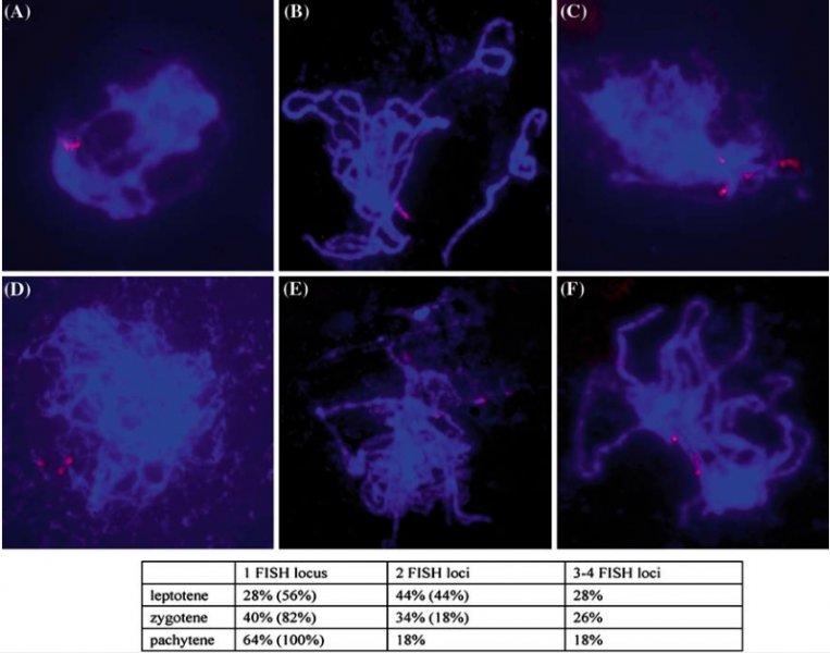
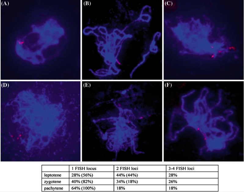
Figure 9. Fluorescence in situ hybridization (FISH) using a probe of 25S rDNA in wild type (A, B) and OsRad21-4-deficient (C–F) plant male meiocytes. The probe was labeled with digoxigenin-labeled dUTP and detected with rhodamin-conjugated anti-digoxigenin antibody (red). Chromosomes were counterstained with DAPI (blue). (A, C) leptotene; (B) pachytene; (D) zygotene; (E) early pachytene; (F) pachytene. The experimental data were summaried and listed below the picture. The data in the bracket were from wild type plants, and the others from the deficient plants. n=50.
File history
Click on a date/time to view the file as it appeared at that time.
| Date/Time | Thumbnail | Dimensions | User | Comment | |
|---|---|---|---|---|---|
| current | 13:37, 22 May 2014 | | 799 × 628 (617 KB) | Mmlin66 (talk | contribs) | Figure 9. Fluorescence in situ hybridization (FISH) using a probe of 25S rDNA in wild type (A, B) and OsRad21-4-deficient (C–F) plant male meiocytes. The probe was labeled with digoxigenin-labeled dUTP and detected with rhodamin-conjugated anti-digoxig |
- You cannot overwrite this file.
File usage
There are no pages that link to this file.