File:Figure6.jpg
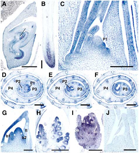
(A) to (I) Antisense probe. (J) Sense probe. (A) Longitudinal section of embryo at 10 d after pollination. sm, SAM; vb, vascular bundle; ra, radicle. (B) Crown root at 1 week after germination. (C) Longitudinal section of 3-week-old shoot apex. (D) to (F) Serial transverse sections of 3-week-old shoot apex. Panels are from the bottom to the top. PLA2 signals are strong in marginal regions. (G) Inflorescence apex at stage In2. Two primary rachis branches (asterisks) are formed. b1 and b2 indicate first and second bracts, respectively. (H) Inflorescence apex at stage In5. Asterisks indicate spikelet primordia. (I) Spikelet at stage Sp6. le, lemma; pa, palea; lo, lodicule; st, stamen. (J) Longitudinal section of shoot apex. No hybridization signals are observed with sense probe. Bars ¼ 500 mm in (A), 250 mm in (B) and (H), 100 mm in (C), (G), and (J), and 150 mm in (D) to (F) and (I).
File history
Click on a date/time to view the file as it appeared at that time.
| Date/Time | Thumbnail | Dimensions | User | Comment | |
|---|---|---|---|---|---|
| current | 03:53, 1 June 2014 | | 475 × 523 (95 KB) | Zhuhuihui (talk | contribs) | (A) to (I) Antisense probe. (J) Sense probe. (A) Longitudinal section of embryo at 10 d after pollination. sm, SAM; vb, vascular bundle; ra, radicle. (B) Crown root at 1 week after germination. (C) Longitudinal section of 3-week-old shoot apex. (D) to (F) |
- You cannot overwrite this file.
File usage
The following page links to this file:


