File:Image2.png
No higher resolution available.
Image2.png (717 × 280 pixels, file size: 238 KB, MIME type: image/png)
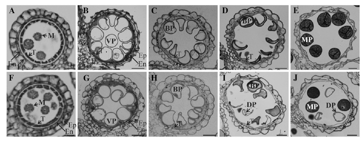
Figure 2. Light microscopy observation of anthers at different developmental stages. Cross sections are shown from segregating wild type (A–E) and OsGT1/osgt1-1 (F–J) at early microspore stage (A and F), vacuolated stage (B and G), mitotic division stage 11a (C and H), mitotic division stage 11b (D and I), and mature pollen stage (E and J). BP, Binuclear pollen; DP, defective pollen; En, endodermis; Ep, epidermis; M, microspore; MP, mature pollen; T, tapetum; VP, vacuolated pollen. Bars = 25 mm.
File history
Click on a date/time to view the file as it appeared at that time.
| Date/Time | Thumbnail | Dimensions | User | Comment | |
|---|---|---|---|---|---|
| current | 05:02, 4 June 2014 | 717 × 280 (238 KB) | Tracy (talk | contribs) | Figure 2. Light microscopy observation of anthers at different developmental stages. Cross sections are shown from segregating wild type (A–E) and OsGT1/osgt1-1 (F–J) at early microspore stage (A and F), vacuolated stage (B and G), mitotic division st |
- You cannot overwrite this file.
File usage
The following page links to this file: