File:Image7.png
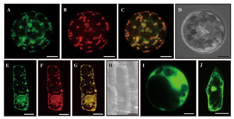
Figure 7. Subcellular localization. A to D, Mesophyll protoplasts prepared from young seedlings were cotransformed with fusion constructs Pubi::OsGT1-GFP and P35S::ManI-RFP. Images observed under GFP channel (A) and RFP channel (B) were merged (C); D is a bright-field image. E to H, Onion epidermal cells were cobombarded with fusion constructs. The GFP channel image (E) and RFP channel image (F) were merged (G); H is a bright-field image. I, GFP image of mesophyll cells xpressing control construct Pubi::GFP. J, GFP image of onion epidermal cell expressing control construct P35S::GFP. Bars = 5 mm for mesophyll protoplasts and 50 mm for onion cells.
File history
Click on a date/time to view the file as it appeared at that time.
| Date/Time | Thumbnail | Dimensions | User | Comment | |
|---|---|---|---|---|---|
| current | 05:14, 4 June 2014 | | 755 × 385 (302 KB) | Tracy (talk | contribs) | Figure 7. Subcellular localization. A to D, Mesophyll protoplasts prepared from young seedlings were cotransformed with fusion constructs Pubi::OsGT1-GFP and P35S::ManI-RFP. Images observed under GFP channel (A) and RFP channel (B) were merged (C); D is a |
- You cannot overwrite this file.
File usage
The following page links to this file: