File:The subcellular localization of OsBAK1 protein in onion epidermis cells.jpg
No higher resolution available.
The_subcellular_localization_of_OsBAK1_protein_in_onion_epidermis_cells.jpg (470 × 367 pixels, file size: 147 KB, MIME type: image/jpeg)
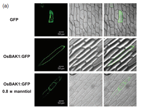
The upper panel shows the 35S::GFP signal alone, which is distributed on the plasma membrane, cytoplasm and nucleus; the middle panel displays the signal from 35S::OsBAK-GFP which is primarily located in the plasma membrane; the lower panel shows 35S::OsBAK-GFP and plasma membrane retracting from the cell wall when onion epidermis cells were treated with 0.8 M mannitol and subsequently plasmolysed. Left line represents green fluorescence signal detected at 488 nm; middle line represents bright field; right line represents the merge of GFP signal and bright field
File history
Click on a date/time to view the file as it appeared at that time.
| Date/Time | Thumbnail | Dimensions | User | Comment | |
|---|---|---|---|---|---|
| current | 08:41, 8 June 2014 | | 470 × 367 (147 KB) | Singing (talk | contribs) | Fig.5 The_subcellular_localization_of_OsBAK1_protein_in_onion_epidermis_cells(The upper panel shows the 35S::GFP signal alone, which is distributed on the plasma membrane, cytoplasm and nucleus; the middle panel displays the signal from 35S::OsBAK-GFP whi |
| 15:50, 5 June 2014 |  | 470 × 367 (147 KB) | Singing (talk | contribs) | The upper panel shows the 35S::GFP signal alone, which is distributed on the plasma membrane, cytoplasm and nucleus; the middle panel displays the signal from 35S::OsBAK-GFP which is primarily located in the plasma membrane; the lower panel shows 35S::OsB |
- You cannot overwrite this file.
File usage
There are no pages that link to this file.


