File list
This special page shows all uploaded files.
| Date | Name | Thumbnail | Size | Description | Versions |
|---|---|---|---|---|---|
| 09:14, 4 June 2014 | Figure 1.The rice hei10-1 mutant phenotype.jpg (file) |  |
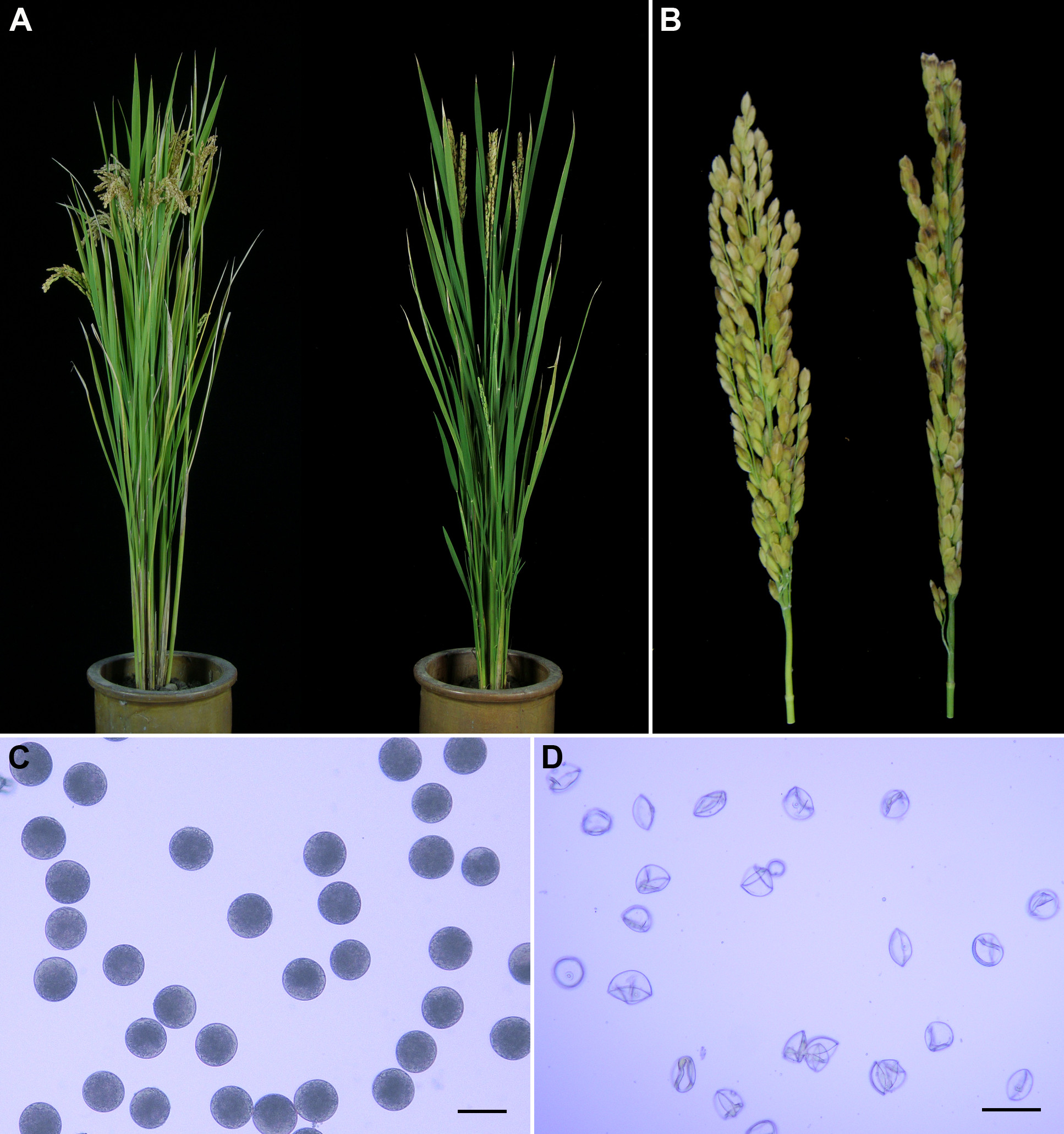
4.34 MB | (A) Comparison of a WT plant (left) and a hei10-1 mutant plant (right). (B) Comparison of a WT panicle (left) and a hei10-1 mutant panicle (right). (C) Fertile pollen grains in a WT plant. (D) Sterile pollen grains in a hei10-1 mutant plant. Scale bars: 5 | 1 |
| 09:50, 4 June 2014 | The rice hei10-1 mutant phenotype.jpg (file) |  |
647 KB | (A) Comparison of a WT plant (left) and a hei10-1 mutant plant (right). (B) Comparison of a WT panicle (left) and a hei10-1 mutant panicle (right). (C) Fertile pollen grains in a WT plant. (D) Sterile pollen grains in a hei10-1 mutant plant. Scale bars: 5 | 2 |
| 09:51, 4 June 2014 | Figure 2.Organization of the HEI10 gene and protein alignment.jpg (file) |  |
18 KB | (A) Schematic representation of HEI10 gene and the location of hei10 mutation. Translated regions are represented by black boxes and untranslated regions are indicated by white boxes. HEI10A and HEI10B indicate long and short transcripts, respectively. d | 1 |
| 10:00, 4 June 2014 | Figure 3.Meiosis in the hei10-1 mutant.jpg (file) |  |
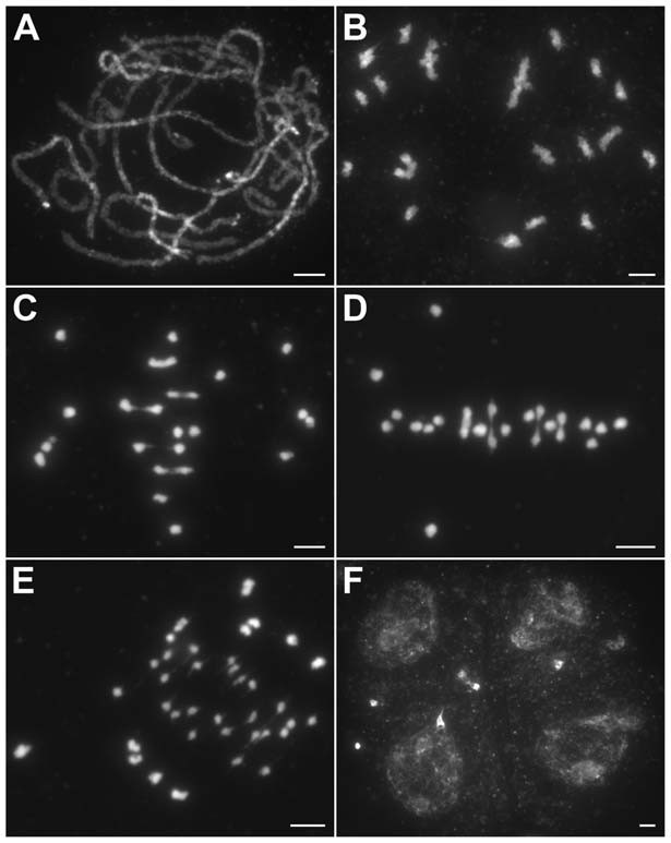
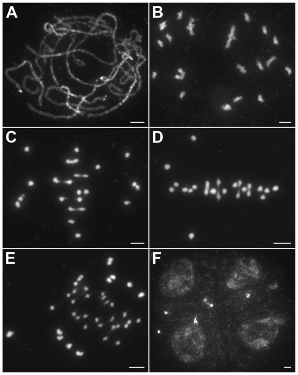
61 KB | (A) Pachytene. (B) Diakinesis with both bivalents and univalents. (C) Metaphase I with five bivalents and fourteen univalents. (D) Metaphase I with four bivalents and sixteen univalents. Thirteen univalents and all bivalents align on the equatorial plate | 1 |
| 10:02, 4 June 2014 | Figure 4. Analysis of the distribution of HEI10 bright foci in WT meiocytes.jpg (file) |  |
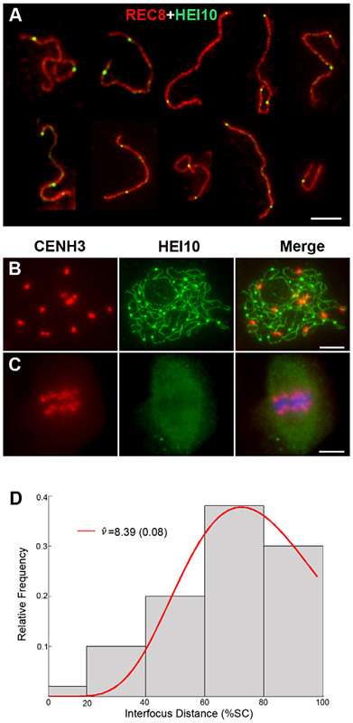
71 KB | (A) Dual immunolocalization of WT scattered chromosomes with REC8 (red) and HEI10 (green) antibodies. Scale bar: 5 mm. (B) Dual immunolocalization of CENH3 and HEI10 at late pachytene. (C) No HEI10 signals were detected at metaphase I, chromosomes are sta | 1 |
| 10:10, 4 June 2014 | Figure111.jpg (file) |  |
647 KB | (A) Comparison of a WT plant (left) and a hei10-1 mutant plant (right). (B) Comparison of a WT panicle (left) and a hei10-1 mutant panicle (right). (C) Fertile pollen grains in a WT plant. (D) Sterile pollen grains in a hei10-1 mutant plant. Scale bars: 5 | 1 |
| 10:10, 4 June 2014 | Figure222.jpg (file) |  |
18 KB | (A) Schematic representation of HEI10 gene and the location of hei10 mutation. Translated regions are represented by black boxes and untranslated regions are indicated by white boxes. HEI10A and HEI10B indicate long and short transcripts, respectively. | 2 |
| 10:11, 4 June 2014 | Figure333.jpg (file) |  |
61 KB | (A) Pachytene. (B) Diakinesis with both bivalents and univalents. (C) Metaphase I with five bivalents and fourteen univalents. (D) Metaphase I with four bivalents and sixteen univalents. Thirteen univalents and all bivalents align on the equatorial plate | 1 |
| 10:12, 4 June 2014 | Figure444.jpg (file) |  |
71 KB | (A) Dual immunolocalization of WT scattered chromosomes with REC8 (red) and HEI10 (green) antibodies. Scale bar: 5 mm. (B) Dual immunolocalization of CENH3 and HEI10 at late pachytene. (C) No HEI10 signals were detected at metaphase I, chromosomes are st | 1 |