Difference between revisions of "File:Interaction of OsBAK1 with OsBRI1 in vivo.jpg"
(Fig.11 Interaction_of_OsBAK1_with_OsBRI1_in_vivo((b) The structure sketch map of OsBAK1 protein. OsBAK1 encoding 624 amino acid residues and including an extracellular domain (amino acid 1–299) and an intracellular domain (amino acid 300–624). The ext) |
(uploaded a new version of "File:Interaction of OsBAK1 with OsBRI1 in vivo.jpg": Fig.11 Interaction_of_OsBAK1_with_OsBRI1_in_vivo(Fig.11 Interaction_of_OsBAK1_with_OsBRI1_in_vivo((b) The structure sketch map of OsBAK1 protein. OsBAK1 encoding) |
(No difference)
| |
Latest revision as of 06:07, 9 June 2014
Fig.11 Interaction_of_OsBAK1_with_OsBRI1_in_vivo((b) The structure sketch map of OsBAK1 protein. OsBAK1 encoding 624 amino acid residues and including an extracellular domain (amino acid 1–299) and an intracellular domain (amino acid 300–624). The extracellular domain of OsBAK1 was named OsBAK1-ECD; the intracellular domain of OsBAK1 was named OsBAK1-ICD; a truncated intracellular domain of OsBAK1 (amino acid 348–624) was named OsBAK1- ICDD. (c) BiFC assay showed the interaction of OsBAK1 and OsBRI1 in vivo. The onion epidermis cells were cotransformated with OsBAK1-YN and YC, OsBRI1-YC and YN, OsBRI1-YC and OsBAK1-YN, OsBRI1-YC and OsBAK1-ECD-YN,OsBRI1-YC and OsBAK1-ICD-YN, and OsBRI1-YC and OsBAK1-ICDD-YN. Left line represents yellow fluorescence signal; right line represents the merge of YFP signal and bright field)
File history
Click on a date/time to view the file as it appeared at that time.
| Date/Time | Thumbnail | Dimensions | User | Comment | |
|---|---|---|---|---|---|
| current | 06:07, 9 June 2014 | 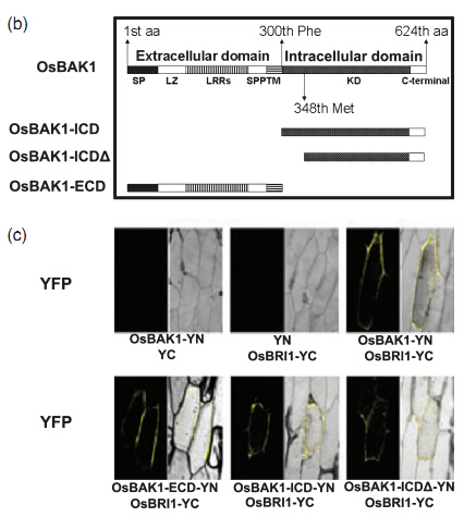 | 426 × 486 (140 KB) | Singing (talk | contribs) | Fig.11 Interaction_of_OsBAK1_with_OsBRI1_in_vivo(Fig.11 Interaction_of_OsBAK1_with_OsBRI1_in_vivo((b) The structure sketch map of OsBAK1 protein. OsBAK1 encoding 624 amino acid residues and including an extracellular domain (amino acid 1–299) and an int |
| 06:02, 9 June 2014 | 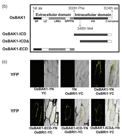 | 426 × 486 (140 KB) | Singing (talk | contribs) | Fig.11 Interaction_of_OsBAK1_with_OsBRI1_in_vivo((b) The structure sketch map of OsBAK1 protein. OsBAK1 encoding 624 amino acid residues and including an extracellular domain (amino acid 1–299) and an intracellular domain (amino acid 300–624). The ext |
- You cannot overwrite this file.
File usage
There are no pages that link to this file.


