Unused files
The following files exist but are not embedded in any page. Please note that other web sites may link to a file with a direct URL, and so may still be listed here despite being in active use.
Showing below up to 50 results in range #501 to #550.
View (previous 50 | next 50) (20 | 50 | 100 | 250 | 500)
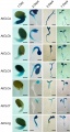 05.jpg 662 × 1,238; 79 KB
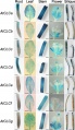 07.jpg 666 × 1,151; 85 KB
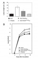 Fig. 5.jpg 384 × 593; 59 KB
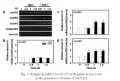 OsGR2 Fig.1.png 1,043 × 702; 194 KB
 Floral phenotype of the sl1 mutant.png 307 × 519; 191 KB
 Scanning election micrographs of sl1 flowers.png 379 × 447; 347 KB
 Molecular cloning of SL 1.png 417 × 475; 74 KB
 Floral morphology of SL1 over-expressors.png 492 × 496; 282 KB
 SL 1 encodes a zinc finger protein with a single C2H2 motif.png 417 × 469; 191 KB
 Regulation of rice floral homeotic genes by SL 1.png 230 × 303; 36 KB
 Possible roles for SL 1 in rice floral organ formation.png 510 × 179; 23 KB
 Identification of the SKC1 QTL and genetic complementation.jpg 148 × 200; 15 KB
 SKC1 promoter–GUS expression pattern in transgenic rice plants.jpg 154 × 200; 15 KB
 Structural model and expression of SKC1..jpg 217 × 200; 21 KB
 Figure 4. Characteristics of FOS1 protein.jpg 500 × 359; 39 KB
 Figure1. RSS3, bHLH, and JAZ Form a Ternary Complex.jpg 717 × 1,544; 150 KB
 1.gif 1,055 × 773; 191 KB
 RSS3 and JAZ9 together Suppress bHLH094-Mediated Gene Activation.jpg 717 × 1,823; 133 KB
 Map-based cloning and characteriztion of TAD1.png 495 × 329; 124 KB
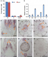 Expression patterns of TAD1.png 427 × 515; 234 KB
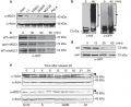 TAD1 targets MOC1 in a cell-cycle phase-dependent manner.png 593 × 483; 122 KB
 The APC C(TAD1) complex-mediated degradation of MOC1.png 645 × 412; 159 KB
 Expression of psbS genes in transgenic plants..png 424 × 512; 88 KB
 Tissue-specific GUS activities in the OsSERK1-GUS rice plants.jpg 558 × 354; 289 KB
 Phylogenetic analysis of SERK proteins in Arabidopsis and rice.jpg 508 × 184; 29 KB
 PNAS-2012-Yang-E1192-200.pdf 0 × 0; 1.53 MB
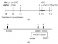 Location of alk.jpg 429 × 326; 13 KB
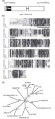 Wjt1.jpg 287 × 623; 40 KB
 Figure 1. Phylogenetic analysis of the XB3 family.png 320 × 414; 33 KB
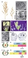 Wjt2.jpg 247 × 514; 26 KB
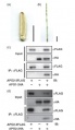 Wjt3.jpg 315 × 544; 18 KB
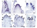 Wjt4.jpg 626 × 480; 58 KB
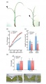 Wjt5.jpg 320 × 581; 22 KB
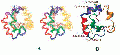 Figure A and B.gif 476 × 220; 27 KB
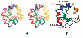 110.gif 476 × 220; 27 KB
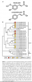 Figure 1xxy.png 263 × 668; 106 KB
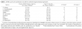 Table 1xxy.png 852 × 304; 76 KB
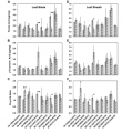 Figure 2xxy.png 512 × 559; 130 KB
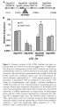 Figure 3xxy.png 354 × 632; 128 KB


_complex-mediated_degradation_of_MOC1.png)


