File:Figure 3. Protein diversity of Ghd7.jpg
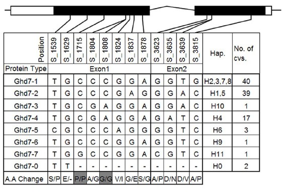
Revision as of 04:13, 31 May 2014 by Lily (talk | contribs) (uploaded a new version of "File:Figure 3. Protein diversity of Ghd7.jpg": The two exons (indicated in black rectangle) and the 59 and 39 UTRs (indicated in white rectangle) of Ghd7 are shown in graphics on the top. The first row indicates th)
No higher resolution available.
Figure_3._Protein_diversity_of_Ghd7.jpg (570 × 383 pixels, file size: 77 KB, MIME type: image/jpeg)
File history
Click on a date/time to view the file as it appeared at that time.
| Date/Time | Thumbnail | Dimensions | User | Comment | |
|---|---|---|---|---|---|
| current | 04:13, 31 May 2014 | | 570 × 383 (77 KB) | Lily (talk | contribs) | The two exons (indicated in black rectangle) and the 59 and 39 UTRs (indicated in white rectangle) of Ghd7 are shown in graphics on the top. The first row indicates the position of the SNPs, the last row reveals the amino acid change. Gray indicates synon |
| 03:32, 31 May 2014 | 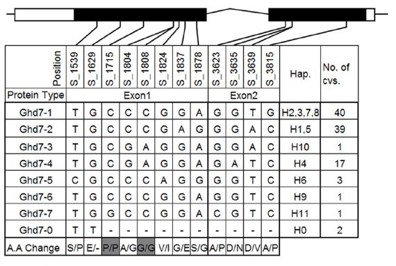 | 570 × 383 (77 KB) | Lily (talk | contribs) | The two exons (indicated in black rectangle) and the 59 and 39 UTRs (indicated in white rectangle) of Ghd7 are shown in graphics on the top. The first row indicates the position of the SNPs, the last row reveals the amino acid change. Gray indicates synon |
- You cannot overwrite this file.
File usage
The following file is a duplicate of this file (more details):
There are no pages that link to this file.


