File:Fig. 2.jpg
Revision as of 06:54, 4 June 2014 by Littlescrew (talk | contribs) (Immunofluorescence microscopy of serial sections of PB-Is obtained from developing rice endosperm at 3 WAF . The numbers at the left of the image denote the order of the sections. The PBs in each section were labeled with antibodies against CysR10 and Cys)
No higher resolution available.
Fig._2.jpg (415 × 597 pixels, file size: 24 KB, MIME type: image/jpeg)
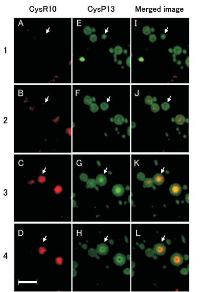
Immunofluorescence microscopy of serial sections of PB-Is obtained from developing rice endosperm at 3 WAF . The numbers at the left of the image denote the order of the sections. The PBs in each section were labeled with antibodies against CysR10 and CysP13. The CysR10–antibody interactions were visualized with rhodamine-conjugated secondary antibodies (A–D), while those for CysP13s were visualized with FITC-conjugated secondary antibodies (E–H). (I–L) Merged images. The arrow indicates the location of the same PB in the serial sections. Bars: 4mm.
File history
Click on a date/time to view the file as it appeared at that time.
| Date/Time | Thumbnail | Dimensions | User | Comment | |
|---|---|---|---|---|---|
| current | 06:54, 4 June 2014 | | 415 × 597 (24 KB) | Littlescrew (talk | contribs) | Immunofluorescence microscopy of serial sections of PB-Is obtained from developing rice endosperm at 3 WAF . The numbers at the left of the image denote the order of the sections. The PBs in each section were labeled with antibodies against CysR10 and Cys |
- You cannot overwrite this file.
File usage
The following page links to this file:


