File:Ma5.jpg
No higher resolution available.
Ma5.jpg (462 × 332 pixels, file size: 222 KB, MIME type: image/jpeg)
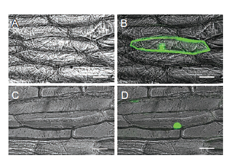
Figure 5 Sub-cellular localization of GW5. (A) and (C) Bright field images of onion epidermal cells. (B) GFP by itself (pCAMBIA 1302 vector) localizes to both cytoplasm and nucleus. (D) GW5-GFP fusion protein is localized only to the nucleus. Scale bars:50 mm.
File history
Click on a date/time to view the file as it appeared at that time.
| Date/Time | Thumbnail | Dimensions | User | Comment | |
|---|---|---|---|---|---|
| current | 13:52, 8 June 2014 | | 462 × 332 (222 KB) | Mafeifei (talk | contribs) | Figure 5 Sub-cellular localization of GW5. (A) and (C) Bright field images of onion epidermal cells. (B) GFP by itself (pCAMBIA 1302 vector) localizes to both cytoplasm and nucleus. (D) GW5-GFP fusion protein is localized only to the nucleus. Scale bars:5 |
- You cannot overwrite this file.
File usage
The following page links to this file:


