File list
This special page shows all uploaded files.
| Date | Name | Thumbnail | Size | Description | Versions |
|---|---|---|---|---|---|
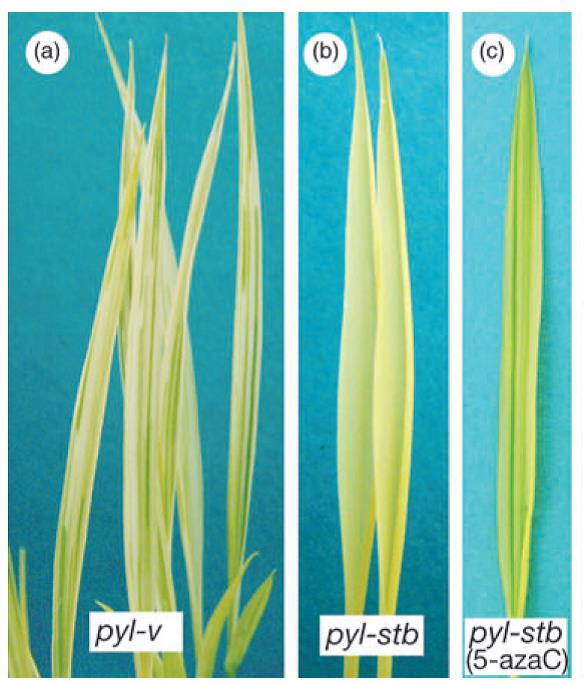
| 13:42, 20 March 2015 | Clpp5.jpg (file) |  |
53 KB | 1 | |
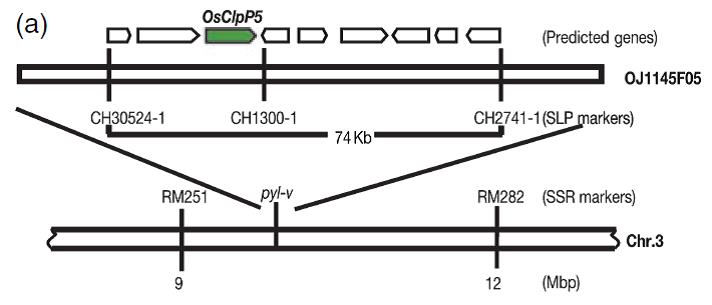
| 13:38, 20 March 2015 | Clpp.jpg (file) |  |
28 KB | 1 | |
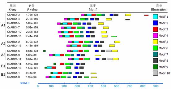
| 09:06, 26 June 2014 | Motif10.jpg (file) |  |
31 KB | Motif composition of rice ABC1 proteins. | 1 |
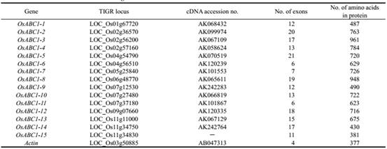
| 04:02, 26 June 2014 | Abc1-table.jpg (file) |  |
20 KB | Table 1. Basic information of the rice ABC1 genes. | 1 |
| 09:33, 10 June 2014 | Oshox-3.jpg (file) |  |
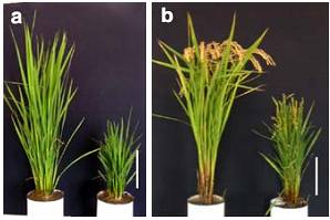
17 KB | Fig. 5 Growth of Oshox4-over expressing and wild type plants treated with GA3 at different concentrations. Wild type (a) and Oshox4-overexpressing plants (b) at four-leaf stage were transferred to hydroponic culture media containing GA3 at the concentrati | 1 |
| 09:32, 10 June 2014 | Oshox-2.jpg (file) |  |
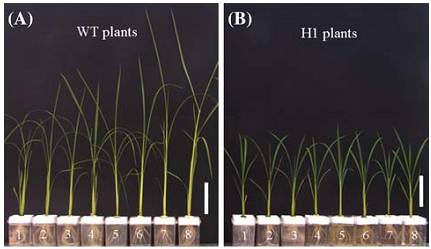
12 KB | Fig. 3 Over-expression ofOshox4induced a semi-dwarf phenotype in transgenic rice. (A) Comparison between wild type and an Oshox4 over-expressing line (H4) at vegetative and reproductive stages. Bars=15 cm. | 1 |
| 09:31, 10 June 2014 | Oshox-1.jpg (file) |  |
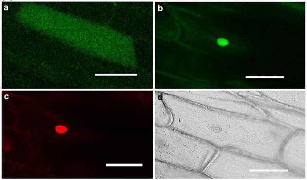
27 KB | Fig.2 (B) Nuclear localization of Oshox4 protein. GFP alone was localized in cytosol of onion skin cells. (a)Oshox4-GFP; Nuclear localised; (c) DAPI staining; (d) a bright light image. Bar=50lm. | 1 |
| 05:08, 6 June 2014 | OsVIT2.jpg (file) |  |
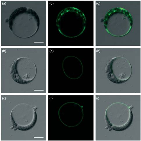
22 KB | Subcellular localization of OsVIT1 and OsVIT2. (a–c) Bright-field images of Arabidopsis protoplasts expressing enhanced green fluorescent protein (EGFP) (a), OsVIT1: EGFP (b) and OsVIT2: EGFP(c). (d–f) Fluorescence images of Arabidopsis protoplasts ex | 1 |
| 06:23, 5 June 2014 | OsPDR92.jpg (file) |  |
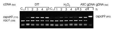
7 KB | Fig. 6.Ospdr9 expression in rice roots in response to DTT, hydrogen peroxide and ascorbic acid. RT-PCR expression patterns of ospdr9 and rac1. Rice seedlings were incubated on growth medium (controls C12 and C13) or on growth medium supplemented with 5 mM | 1 |
| 06:22, 5 June 2014 | OsPDR91.jpg (file) |  |
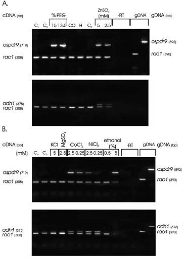
21 KB | Fig. 2. Early ospdr9 expression in rice roots in response to various stresses. RT-PCR products of ospdr9 and rac1 (upper panels) and adh1 and rac1 (lower panels) were separated on 1.1% agarose gels and stained with ethidium bromide.3RT lanes show typical | 1 |
| 14:26, 4 June 2014 | Crp10.jpg (file) |  |
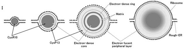
15 KB | Fig. 5 (I) Schematic images of PB-I structure during rice endosperm development. | 1 |
| 06:54, 4 June 2014 | Fig. 2.jpg (file) |  |
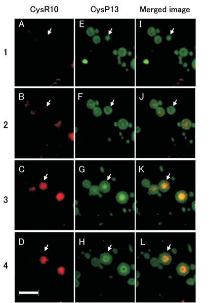
24 KB | Immunofluorescence microscopy of serial sections of PB-Is obtained from developing rice endosperm at 3 WAF . The numbers at the left of the image denote the order of the sections. The PBs in each section were labeled with antibodies against CysR10 and Cys | 1 |